Forensic Imaging After remaining unchanged for nearly two centuries, the world of autopsy and forensic medicine has experienced an unprecedented upheaval with the introduction of imaging.
“I shot him in the head to put an end to his suffering.” The sentence is short, but for the doctors at the University Centre of Legal Medicine in Lausanne (CURML) it is yet another enigma to solve: is the person who did the shooting telling the truth? Or was his real intention to get rid of the other person? Silke Grabherr, head of the Forensic Imaging Unit, has already come up against these situations hundreds of times. “Our job is not only to find the causes of death,” she says. “The witness statements must be systematically compared with the reality expressed by the body. And to do this, we cannot overlook even the tiniest injury.” The specialist and her team have therefore developed a unique technique, which combines systematically scanning all bodies that arrive at the morgue and – if necessary – carrying out an angiogram using a revolutionary technique. Explanations in six cases.
La mort immortalisée from IN VIVO on Vimeo.
Alejandro Dominguez explains what this new technology changed for the imaging specialists.
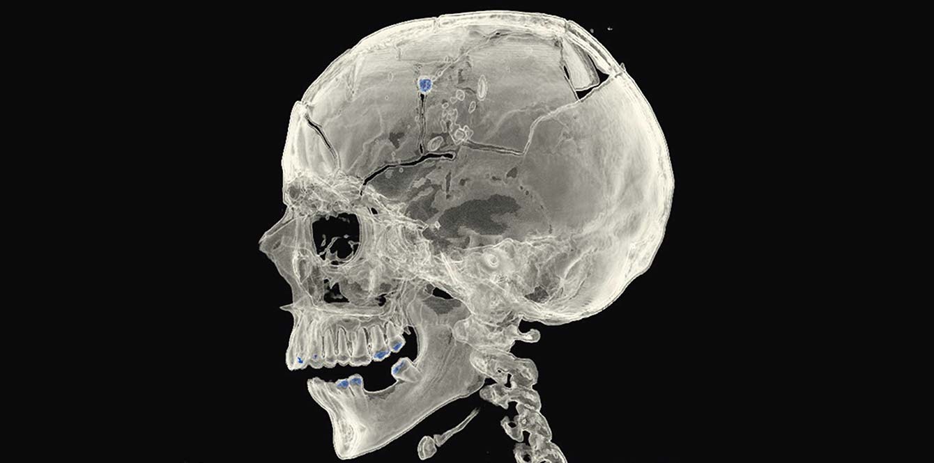
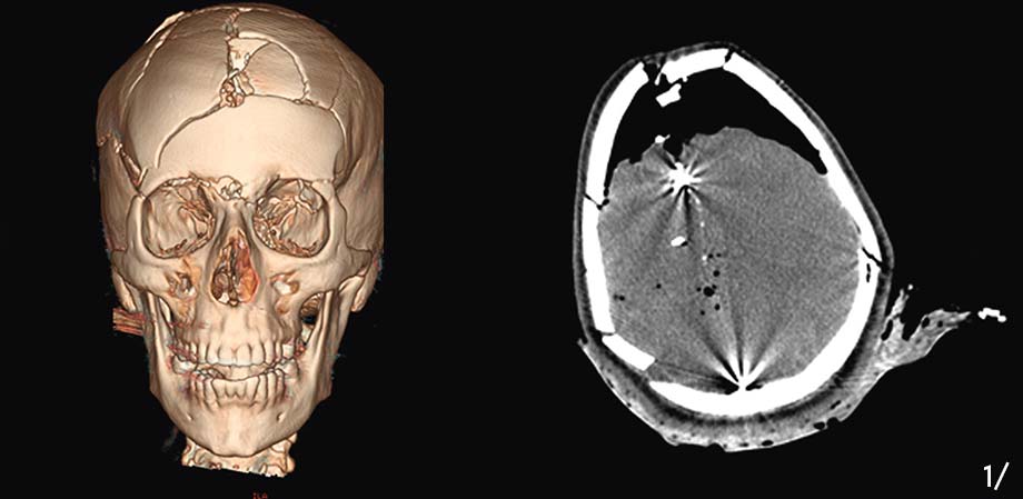
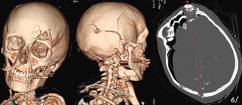
The three images opposite show the skull and brain of the same person, who died from a gunshot wound. The bullet can be seen as the bright spot in the frontal section of the brain (left). The cadaver is one of the 500 to 600 cases treated every year by forensic medicine specialists in the Lausanne region.
If a death is classified as “suspicious” or “violent”, the body is brought to the Centre for Forensic Medicine for a scan and external examination. It is then up to the prosecutor involved in the case to decide whether the specialists should perform an autopsy and, potentially, an angiography (see image above). This medical imaging technique is used to visualise the blood vessels.
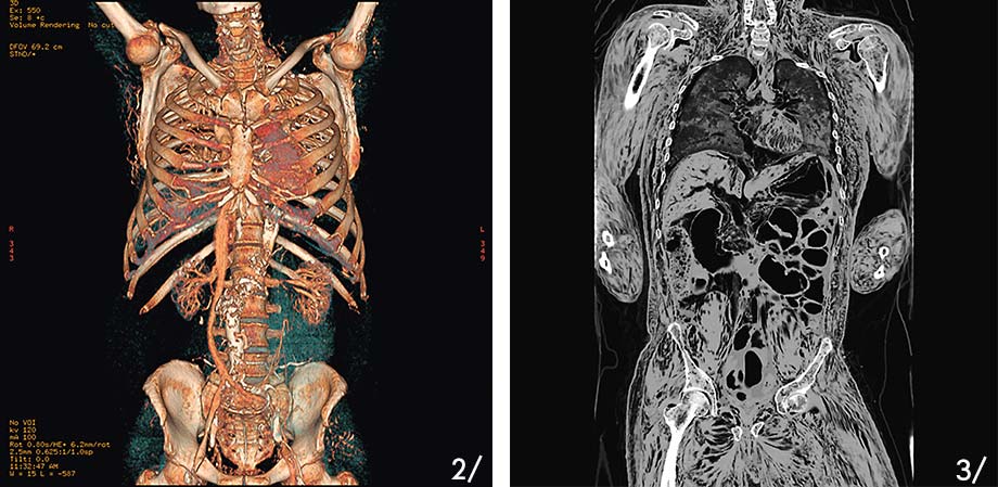
For the novice radiologist, images of a dead body are a shocking spectacle. “Six to eight hours after death, air bubbles can already form inside the body (seen as black spots in the right-hand image). If the specialist does not know that the person is dead, they will diagnose it as an embolism or other pathology,” says Silke Grabherr. “It would be understandable, but wrong. In a cadaver, these bubbles can come from the blood settling with respect to the position of the body or from distortions caused by rigor mortis.”
The person pictured above, who has a major skull fracture, was the victim of a fall at their place of work. The coroner can therefore determine the precise point of impact.
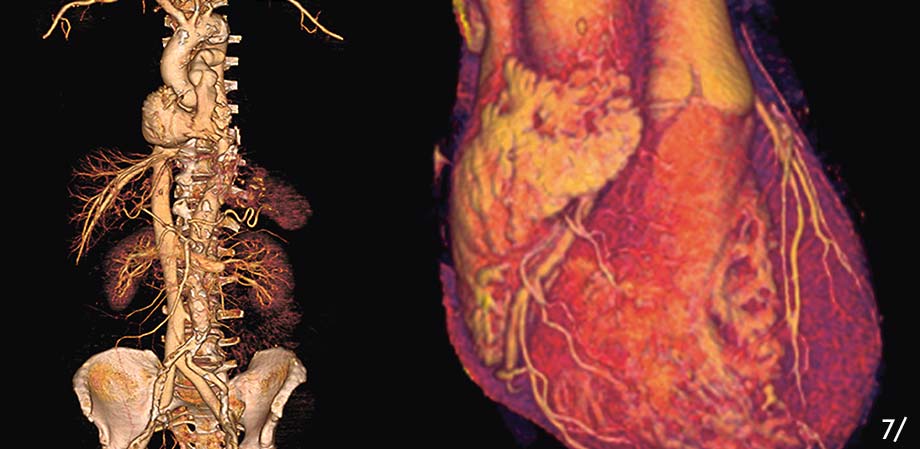
When we die, our heart stops beating. We all know that, but this presents a practical problem. The contrast dye injected, which is required for an angiography, cannot flow. No more heartbeat, no more natural blood circulation and therefore no more flow of the contrast dye through the blood vessels. “To work around the problem, we had to find a way for the dye to circulate,” says Silke Grabherr. “It took several years of research to find the ideal solution that would provide images used to reproduce the blood vessels exactly (see image above).”
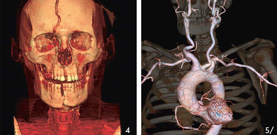
Below is a second 3D reconstitution image of the skull of a victim of a gunshot wound to the head. Thanks to the imaging technology, the projectile’s trajectory was reproduced (see image opposite) without moving the bone or bullet fragments inside the head. Seeing inside the body without opening it is one of the great advantages of forensic imaging. As well, there is a great savings of time: an angiography takes two hours at most, whereas a traditional autopsy of the body of a knife-wound victim can take up to 10 hours.
This reconstitution of the vessels in the chest and abdomen would never have been seen in a traditional autopsy. Today, specialists from numerous countries come for training in forensic imaging techniques developed in Lausanne. “Some countries are turning to this non-invasive method for cultural reasons,” says Silke Grabherr. “Especially in Arab countries, where opening the bodyof a deceased person poses a religious problem.”
Our job is not only to find the causes of death. The witness statements must be systematically compared with the reality expressed by the body. And to do this, we cannot overlook even the tiniest injury.