Every year, amniotic membrane is used as a dressing to treat dozens of patients suffering from eye conditions. A closer look at its unparalleled curative properties.
Human placenta has amazing capacities. This ephemeral organ connects the embryo to the uterine wall to supply the nutrients that the foetus needs to grow. It also turns out to be an effective biological bandage. Boasting anti-inflammatory and anti-bacterial properties, amniotic membrane contains growth factors that accelerate healing, offering benefits that surpass all other treatments available on the market. First of all, it can be used as a graft in eye surgery. “The placenta has the strong advantage of having no antigens, which significantly reduces the risk of rejection,” says Dr Kattayoon Hashemi, a cornea and refractive surgery expert at the Jules-Gonin Ophtalmic Hospital in Lausanne.
But how can an organ be transformed into a bandage? It all starts with a scheduled C-section. “It is crucial for the placenta to remain in a sterile environment, which means it cannot be removed during natural childbirth,” says Dr Michaël Nicolas, head of the Eye Bank at the Ophtalmic Hospital. At Lausanne University Hospital, pregnant women are given the choice to donate their placenta about three weeks before their due date.
“People, even healthcare providers, are relatively unfamiliar with the procedure. A single placenta can produce 20 to 30 bandages, and we need between 35 and 40 grafts per year. Two placentas a year are more than enough,” says Dr Nicolas.
He is also in charge of providing assistant physicians with the guidelines for this exceptional procedure.
Once the placenta is removed, the delivery room doctor places it in a recipient containing antibiotics. “We bring all the required materials the day before to avoid having to go into the delivery room,” Dr Nicolas explains. Still considered by many as waste, this temporary organ becomes a precious good that is handled with the utmost care.
The Eye Bank technicians start by washing the placenta by hand. The organ is tested for bacterial contamination in each phase of the washing process. Then the chorion, the outermost layer of the placenta, is separated, leaving only the amniotic membrane. The entire procedure is performed in a tissue culture hood to maintain a sterile environment. Next comes the cutting process. “Surgeons need different-sized tissue fragments. Sometimes they have to fold the membrane to fill large or deep eye wounds. We produce bandages measuring between two and five square centimetres,” Mr Nicolas says. The grafts are then frozen and can be used within up to two years. Before the transplant, the membrane undergoes a final wash and bacterial test to prepare it for use in the operating room.
This type of bandage can be used in many ways: to treat eye ulcers, ocular surface trauma due to injury or chemical burns, corneal perforation, pterygium (tissue growth from the conjunctiva that invades the cornea), or to prepare the eye for a cornea transplant.
Literally grafted onto the surface of the eye with sutures or surgical glue, the membrane is incorporated into the eyeball or affixed onto the surface like a contact lens.
It is never removed. Drops or contact lenses have more or less the same properties as these biological bandages, but contact lenses increase the risk of infection and drops increase the risk of developing cataracts or glaucoma. Not to mention the high cost of these treatments. Tested for the first time in the 1940s, amniotic membrane transplants have been in use in Lausanne for about 15 years and show no sign of slowing.
On 3 January 1843, cataract patient Elisabeth de Cerjat, ophthalmologist Frédéric Recordon, and philanthropist and banker William Haldimand met in Lausanne. Despite having seemingly little in common, these three people came up with the idea of creating a hospital to treat eye conditions and an institute to teach and train young visually-impaired patients. In the 1970s, the institute was officially named the Jules-Gonin Ophtalmic Hospital after the famous ophthalmologist from the Canton of Vaud. Today, more than 600 people work at the Jules-Gonin Opthalmic Hospital, but also at the Learning Centre for the visually impaired, the Recordon care facility in Lausanne and the Clair-Soleil care facility in Écublens. The Jules-Gonin Ophtalmic Hospital remains the only establishment of its kind in Switzerland.

A laboratory where biologists and technicians remove, transport, preserve, assess and distribute eye tissue, such as corneas, from donors.
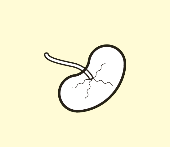
The placenta develops to supply the foetus with nutrients during pregnancy and generally ends its existence in a hospital incinerator. In recent years, the organ has regained its prestige earned in the Middle Ages, when it was believed to hold magical powers. These days, it is used in medicine, and certain women use it as a homeopathic remedy to fight post-partum depression.
These macromolecules are what induce an immune response by antibodies. This response occurs to protect the organism from “foreign” substances but must be prevented in the case of a transplant.